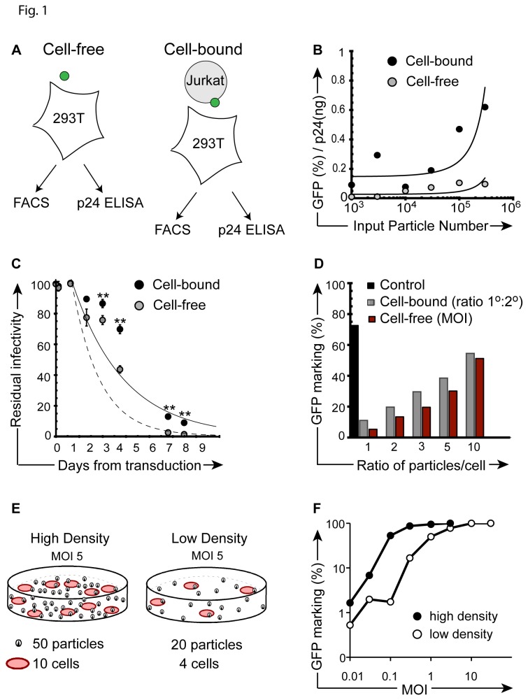
Figure 1. Enhanced gene transfer after direct cell-cell contact of 1o and 2o targets.
(A) Experimental design, illustrating cell-free (left) and cell-bound (right) transduction strategies. (B) Ratio of relative infectivity (% GFP / ng p24) after 24-hour direct cell-free transduction versus transduction by (Jurkat) cell-bound particles following co-culture. The y-axis ratio reflects GFP marking (FACS) per ng input vector (p24 ELISA) at increasing vector particle numbers (x-axis), corresponding to MOI 0.01-3. Refer to Methods section for detailed procedure. (C) Residual infectivity of cell surface bound versus cell-free particles. Particles were kept at 37oC and identical aliquots (MOI 1, based on vector titer) were removed at indicated time points for transduction culture of 1 x10⁵ 293T cells (cell free, black circles). For the cell-bound conditions, the same number of vector particles were arrested on the surface of 1o target Jurkat cells (MOI 1) at 4oC, before shift to 37oC. Aliquots of 1 x10⁵ cells were subsequently placed in coculture with 293T 2o targets at the indicated time-points. A 2-tailed paired Student’s t test was performed; p values ≤ 0.01 are indicated by double asterisks. (D) Relative GFP marking in secondary 293T cells after direct co-culture (24 hours) by escalating the number of GFP vector exposed Jurkat cells (1o, CD45+) to 293T (2o, CD45-) cells at stable MOI of 1 (ratio, gray bars), or by escalating MOI during exposure of primary cells, with matched 1:1 cell numbers (MOI, red bars). Direct transduction of 293T cells with cell-free vector (black bar, control). (E) Illustration of experimental design used in (F). (F) Transduction of Jurkat cells under high cell density transduction (1x10⁶ cells per ml, black circles) or low cell density (1x10⁵ cells per ml, open circles) transduction conditions over a range of MOI. Cells were transduced in the presence of 4 µg/ml protamine sulfate. Following a 3-hour transduction at 37°C, cells were washed, placed back in culture at 37°C, and flow cytometry was performed 72 hours later. All experiments were repeated with similar results.

