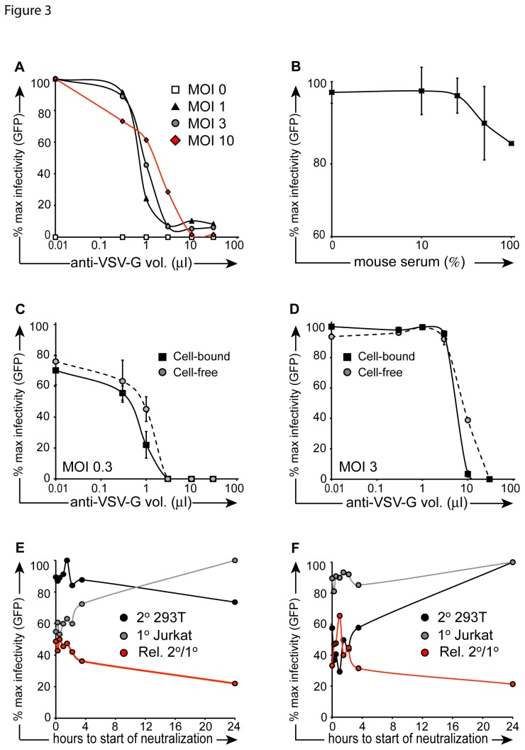
Figure 3. Neutralization kinetics of cell-free vector.
(A) Vector particles were incubated in escalating concentrations of anti-VSV-G neutralizing antibody for 1 hour at 4°C in the presence of 4 µg/ml protamine sulfate. Particles were then placed on 293T cells for overnight transduction at 37°C. Cells were washed after 24 hours, and FACS was performed 48 hours later. (B) Experiment was performed as described in (A) with the exception that mouse serum was used instead of VSV-G neutralizing antibody and the neutralizing incubation was performed at 37°C for the purpose of minimizing cell death resulting from serum incubation (CD). In the cell-bound condition (black squares), 1x10⁵ prechilled Jurkat cells were exposed to vector (C: MOI 0.3; D: MOI 3) for 1 hour at 4°C in the presence of 4 µg/ml protamine sulfate, then placed in co-culture with 2.5x10⁴ pre-plated 293T cells at 37°C. Cell-free samples (gray circles) were prepared in parallel, in the absence of Jurkat cells. Escalating concentrations of anti-VSV-G neutralizing antibody were added to co-cultures and incubataed overnight at 37°C. Cells were then washed, and FACS was performed 48 hours later (EF). 1x10⁵ prechilled Jurkat cells were exposed to vector (MOI 3) for 1 hour at 4°C, then placed in co-culture with 2.5x10⁴ pre-plated 293T cells at 37°C. At serial time points (x-axis), 0.5% anti-VSV-G neutralizing antibody (E) or 10% mouse serum (F) was added to the transduction culture. Target 293T cells were washed 24 hours after vector exposure, and FACS was performed 48 hours later. CD45-APC antibody was used to exclude primary Jurkat cells (gray circles) from secondary target 293T cells (black circles). Red line depicts % 293T GFP+ events/ % Jurkat GFP+ events.

