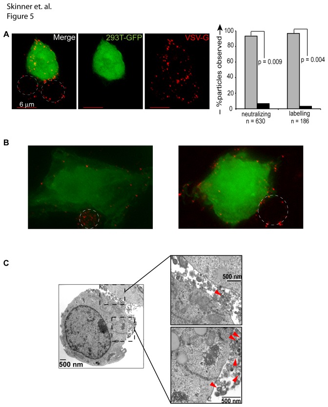
Figure 5. Vector particles do not consistently polarize to areas of cell-cell contact.
(A) Jurkat cells were exposed to mCherryvpr vector (red) for 1 hour at 4°C, cells were washed and placed in co-culture with GFP-expressing 293T cells (green) that were pre-plated on glass cover slips. Cells were washed with PBS, stained with anti-VSV-G antibody (shown) or neutralizing anti-VSV-G antibody (not shown), fixed with 4% paraformaldehyde, and then stained with Alexa Fluor 647 anti-rabbit secondary antibody (magenta). The percentage of single or aggregated particles was enumerated in cells treated with neutralizing versus labeling VSV-G antibody. Statistical significance was determined via a 2-tailed Students t test assuming unequal variance. (B) A representative image was acquired as described in (A) and used to generate a projection image of 13 z-stacks in softWoRx Explorer. (C) 1x10⁶ Jurkat cells were transduced overnight at 37°C (MOI 25). Cells were washed twice in the morning, pelleted, fixed for 1 hour in Karnovsky’s fixative, and prepared for electron microscopy. Panel on left is magnified 14,000x; the right top panel is magnified 36,000x. Red arrowheads highlight vector particles.

