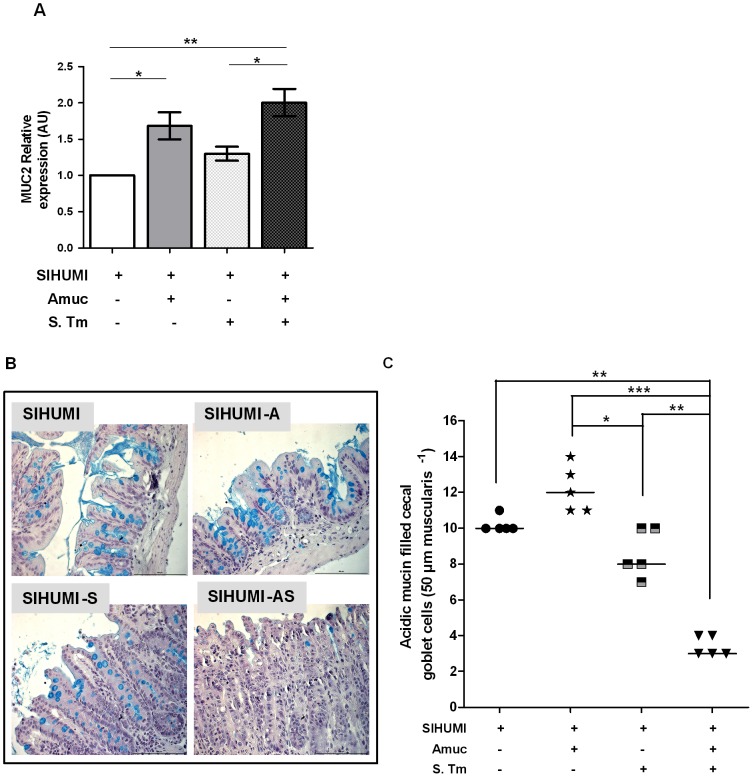
Figure 7. SIHUMI mice with both A. muciniphila and S. Typhimurium display increased MUC2 mRNA levels (A) and reduced numbers of mucin filled goblet cells (B and C).
(A) mRNA was extracted from cecum mucosa of mice belonging to either one of four groups: SIHUMI, SIHUMI-A, SIHUMI-S and SIHUMI-AS. MUC2 mRNA from cecum mucosa was converted to cDNA and expression levels were quantified using real-time PCR (see materials and methods). SIHUMI-A and SIHUMI-AS mice showed significantly higher MUC2 gene expression compared to the other two groups, harboring no A. muciniphila. Data are expressed as mean±standard error. n = 6 per group. *P<0.05, **P<0.01, ***P<0.001. Amuc: A. muciniphila; S. Tm: S. Typhimurium. (B) Formalin fixed cecal tissue sections from SIHUMI, SIHUMI-A, SIHUMI-S and SIHUMI-AS mice were stained with alcian blue (pH-2.5) and haematoxylin. Images are representative of 5 mice per group. Magnification 400-fold. SIHUMI-AS mice display the lowest number of positively stained mucin-filled goblet cells compared to the other three groups. The bar represents 100 µm. (C) Quantitative analysis of the number of acidic mucin-filled goblet cells (blue) enumerated in cecal tissue sections from SIHUMI, SIHUMI-A, SIHUMI-S and SIHUMI-AS mice for a 50 µm stretch of lamina muscularis corresponding to approximately 30 cecal crypts per section. Two sections per mouse were analyzed. The number of cecal mucin filled goblet cells was elevated when A. muciniphila was present (SIHUMI-A) but the concomitant presence of S. Typhimurium (SIHUMI-AS) resulted in the lowest number of mucin filled goblet cells of gnotobiotic SIHUMI mice compared to the other mouse groups. Data are expressed as mean±standard error. n = 5 mice. *P<0.05, **P<0.01, ***P<0.001. Amuc: A. muciniphila; S. Tm: S. Typhimurium.

