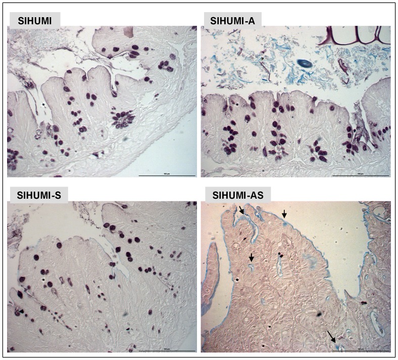
Figure 8. SIHUMI mice colonized with both A. muciniphila and S. Typhimurium display reduced mucus sulphation.
Formalin fixed thin sections (4 µm) of cecal tissue of mice belonging to either one of four groups: SIHUMI, SIHUMI-A, SIHUMI-S and SIHUMI-AS (see Figure. 1) were stained with high iron diamine (HID)/AB at pH-2.5 and subsequently analyzed. Brown color indicates sulphated mucins while blue color indicates sialylated mucins. SIHUMI-AS mice display few sulphated mucins compared to the other mouse groups. Magnification 400×. Bars indicate 100 µm.

