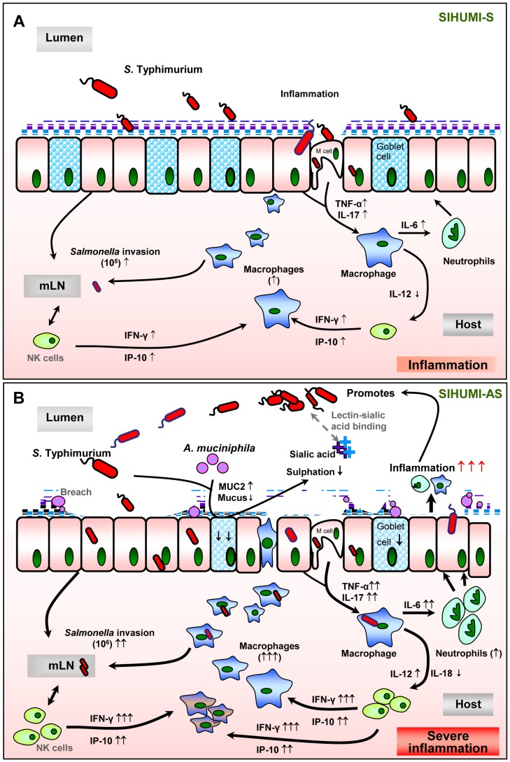
Figure 9. Hypothetical Scheme.
The presence of A. muciniphila, leads to the exacerbation of S. Typhimurium-induced intestinal inflammation. We propose that the presence of A. muciniphila causes changes in mucin composition and production, which in turn facilitates the invasion of S. Typhimurium into the host. Increased inflammatory status was characterized by increased pro-inflammatory cytokines, increased macrophage infiltration and invasion of the pathogen into the lymph nodes, reduced number of mucin-filled goblet cells in SIHUMI-AS mice (B) compared to SIHUMI-S mice (A). Our data suggests that in the presence of both A. muciniphila and S. Typhimurium, mucus sulphation is diminished and this may facilitate the access of S. Typhimurium to sialic acid in mucus. Sialic acid may serve as a substrate and adhesion site for S. Typhimurium in the gut [56], [57]. Increased gene expression of IFN-γ and IP-10 indicate an increased NK-cell recruitment. mLN - mesenteric lymph nodes, NK- Natural killer cells. (↑ increased; ↓ decreased; grey dotted line: assumed processes including lectin-sialic acid binding [56], M-cells for pathogen transit [43], [58], [59]; black line: supported by data of the present study).

