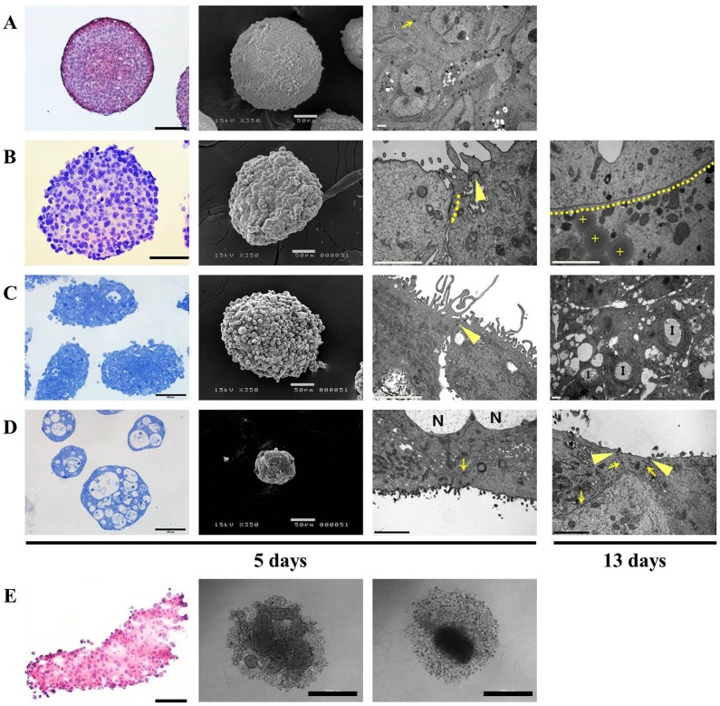
Figure 3. Morphology and histological examination of tumor spheroids (TS) cultured in concave microwell 600 or in 96 well plates.
Representative images of H&E stained paraffin sections or toluidine blue stained semi-thin sections, SEM and TEM images of HT-29 (A), Panc-1 (B), Aspc-1 (C), and Capan-2 (D) spheroids cultured in concave microwell 600 plates for 5 and 13 days. Aggregates of Panc-1, Aspc-1, and Capan-2 cells formed in agarose-coated 96 well plates shown as an H&E stained paraffin section or bright field images (E). Arrow: desmosome; dotted lines: gap junctions; arrow head: tight junction; cross: lipid droplet; I: invagination structure; N: necrotic regions. The scale bars indicate 100 μm, 50 μm, 2 μm and 500 μm, in H&E or toluidine blue stained, SEM, TEM and bright-field images, respectively.

