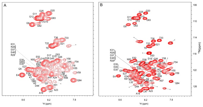
Figure 1. Pup is disordered inside E. coli.
A. In-cell 1H{15N}-HSQC spectrum of [U-15N] Pup-GGQ. B. In vitro 1H{15N}-HSQC spectrum of [U-15N] Pup-GGQ. The comparatively high viscosity of the cytosol, relative to that of the cell lysate, results in a lower rate of tumbling and consequent peak broadening in the in-cell spectrum. Conversely, the lower viscosity of the cell lysate allows faster tumbling and generates sharper resonances in the in vitro spectrum. For quantitation of changes in chemical shifts and peak intensities see Figure S1 . Asterisks denote NMR peaks originating from small metabolites.

