
Figure 1. Development of endometriotic lesions in fat-1 and wild type mice.
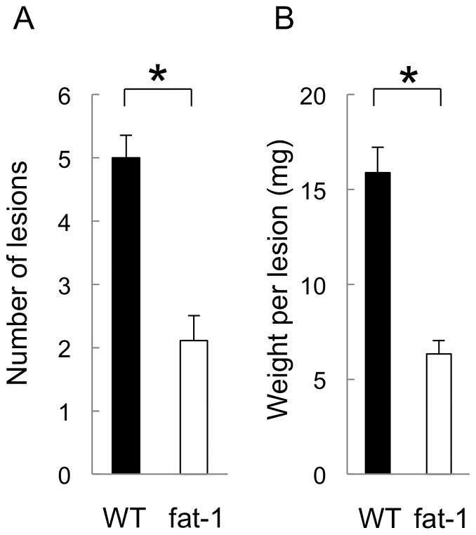
A cystic mass was histologically confirmed as an endometriotic lesion. (A) The number of lesions was counted macroscopically. (B) All masses were resected. The weight (mg) per lesion was measured. These data were compared between the fat-1 and wild type (WT) mice (n = 10 in each group). Mean values with standard deviations are presented. Asterisks indicate those comparisons (fat-1 vs. wild type mice) with statistical significance (p<0.05).