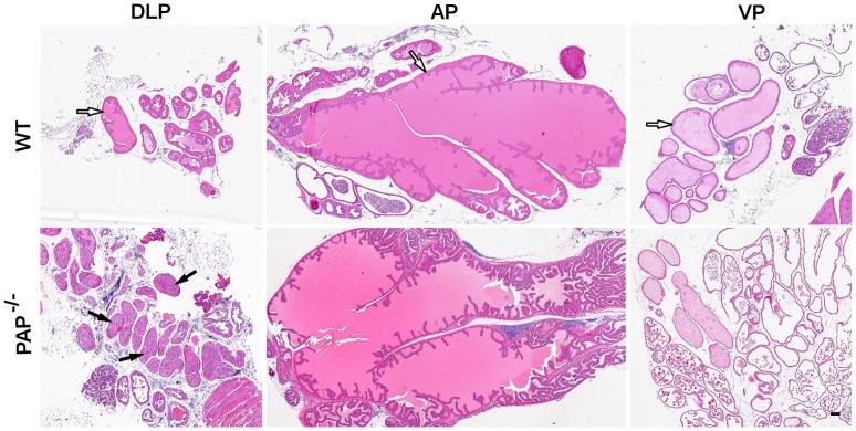
Figure 1. DLP lobeexhibits the primary changes in the PAP−/− mouse prostate.
The panels show an overview of the 12-old mice prostate dissected lobes. The DLP, AP and VP lobes were dissected from WT and PAP−/− mouse. The monolayer epithelium (white arrows) is seen in all the lobes of the WT mouse, whereas in the PAP−/− mouse an increased amount of cells is present in the lumen of the DLP lobe (black arrows). The AP and VP of PAP−/− mouse show no significant changes. Scale bars: 100 µm.

