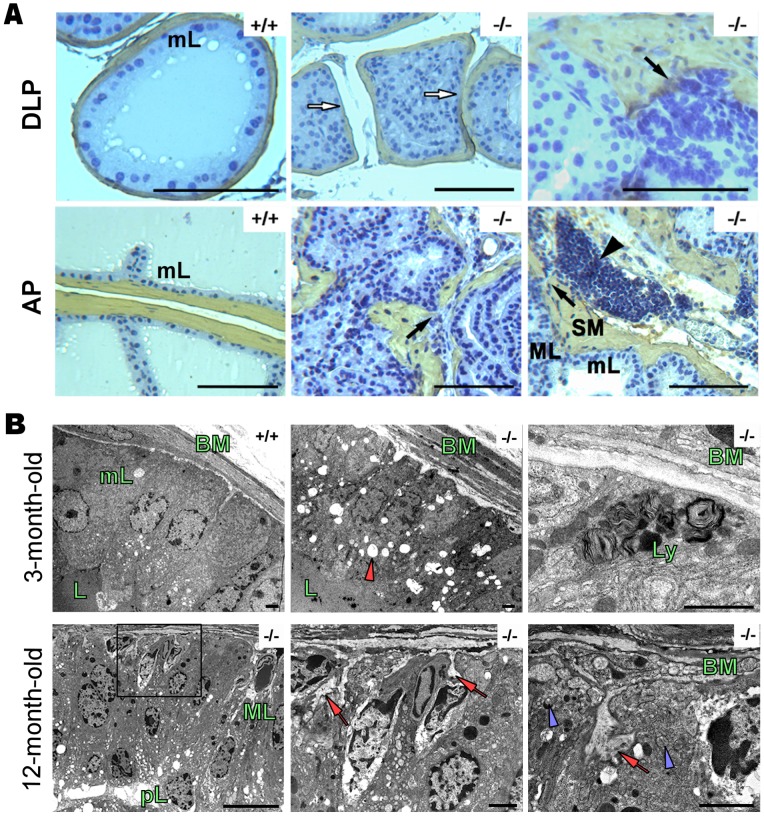
Figure 3. The prostate adenocarcinoma in PAP−/− mice is also detected by immunohistochemistry and electronmicroscopy.
A, smooth muscle actin (SMA) immunohistochemistry in 12 month-old mice. Monolayer epithelium (mL) and open lumen in PAP+/+ DLP. White arrows show the broken fibromuscular sheath (SM, smooth muscle) and bulging of epithelial cells to the stroma. Prostate adenocarcinoma (black arrows) is present in AP and DLP, showing a multilayer epithelium (ML) and inflammatory cells (black arrowhead) spreading in neighboring areas. Scale bars: 100 µm. (n = 4, per group). B, ultrastructural changes in 3 month-old and 12 month-old PAP−/− mouse DLPs. Monolayer epithelium, regular basement membrane (BM) and apical secretion are clearly seen in PAP+/+ mouse DLPs. 3 month-old PAP−/− DLPs show irregular BM and numerous apical vacuoles (red arrow head), as well the presence of basal lysosomes (Ly). In 12 month-old PAP−/− mouse DLPs, the epithelium has transformed to a multilayer epithelium containing hyperchromatic nuclei with multiple nucleoli. Pseudolumens (pL) have formed as a result of the growing and fusion of the epithelium. Invaginations of BM (red arrows) into the epithelium and numerous vesicles in the basal side of the cells (blue arrow heads) were additional signs of the transformation in the cells. Scale bars: 2000 nm (n = 4, per group).

