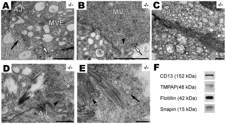
Figure 4. PAP−/− mouse DLPs release exosomal-like microvesicles.
A, electronmicroscopy images show the presence of electron-dense (white arrow) and electron-lucent (black arrow) microvesicles (∼30 to 80 nm) in the lumen of the acini, and MVE containing microvesicles in the apical part of the cell. Scale bar: 1,000 nm. B, numerous microvesicles are present in the apical region of PAP−/− DLP and secreted into the lumen, decreased amount of microvilli is observed (black arrowheads) (scale bar: 1,000 nm). C, microvesicles are secreted into basolateral intercellular space of PAP−/− DLP (scale bar: 2000 nm). D, lamellar body-like structures (*) are inside the epithelial cell (scale bar: 500 nm) and E, released into the lumen (*). Scale bar: 200 nm. F, TMPAP and snapin are also present in exosomes. Immunoblots of exosomes isolated from TMPAP/LNCaP cell culture medium. Flotillin and CD13 were used as exosomal and prostasomal marker respectively.

