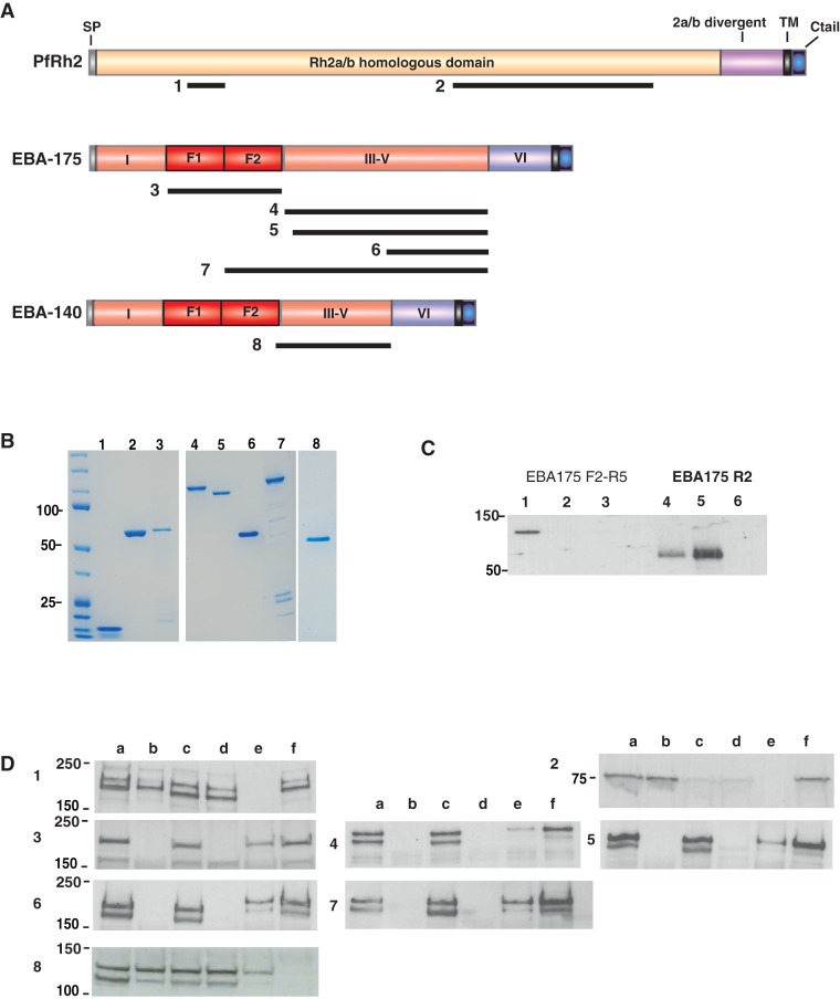
Figure 1. Antibodies raised in rabbits against recombinant P. falciparum invasion ligands specifically recognize native parasite antigens.
A: Schematic of P. falciparum invasion ligands used to generate antigens for immunization. Black bars below the schematic structures indicate the regions of expressed recombinant proteins. Numbers indicate the lanes on the coomassie gel shown in fig. 1B. B: Antigenic fragments of invasion-related proteins were expressed and purified for rabbit immunization. Immunogens are shown here on SDS-PAGE. (1) Rh2 N-terminal fragment (2) Rh2 C-terminal fragment (3) EBA-175 Region 2 (4) EBA-175 Region 3-5 3D7 (5) EBA-175 RIII-V W2mef (6) EBA-175 RIV-V (7) EBA-175 F2-R5 (8) EBA-140 RIII-V. Proteins were stained with coomassie blue. C: Recombinant Region II of EBA-175 binds to erythrocytes in a sialic-dependent manner. Recombinant proteins EBA-175 F2-RV and RII were tested for binding to erythrocytes. Lanes 1 & 4 show protein pre-assay; 2 & 5, binding to untreated erythrocytes; Lanes 3 & 6, binding to neuraminidase-treated erythrocytes. D: Rabbit antibodies raised to recombinant antigens specifically recognize native parasite proteins. 1–8 correspond with antigens as described in Fig. 1A. Lanes as follows: 3D7 wild type (a), 3D7Δ175 KO (b), W2mef (c), W2mefΔ175 KO (d), FCR3 wild type (e), 3D7Δ140 KO (f).

