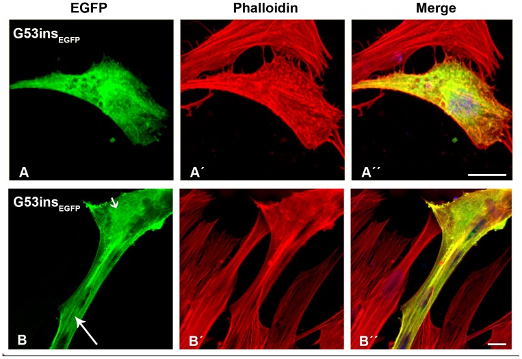
Figure 4. The expression of G53ins-β-TMEGFP in human cells.
The G53ins-β-TMEGFP mutant was transfected in human (A) myoblasts and (B) myotubes and labeled with TRITC-phalloidin (red) and DAPI (blue) to highlight cell nuclei. The G53ins-β-TMEGFP mutant produced delocalisation and endogenous actin aggregates in human myoblasts, labeled with phalloidin (A–A”). The cells transfected with the G53ins-β-TM mutant differentiated into myotubes in an advanced, developed state, identified by their elongated shape and multiple nuclei (B–B”). Transfected myotubes showed good integration of the mutant TM into sarcomeric structures (B; long arrow). The G53ins mutant produced diffuse cytoplasmic labeling at the far end of the myotubes (B; short arrow). Confocal microscopy was performed using a Zeiss LSM 510 Meta confocal microscope or an LSM 700 inverted Axio Observer.Z1 microscope. Scale bar = 10 µm.

