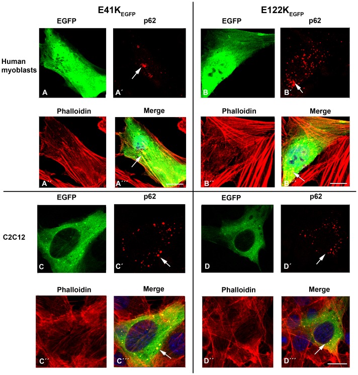
Figure 13. Immunofluorescent labeling of p62 in human myoblasts and C2C12 transfected with E41K-β-TMEGFP and E122K-β-TMEGFP constructs.
The cells were labeled with TRITC-phalloidin (red) and DAPI (blue) to highlight cell nuclei. (A–A’”) The co-localisation of p62 (red, A’; arrow) in aggregates induced by the transfection of E41K-β-TMEGFP in human myoblasts (A and A’”; arrows). (B–B’”) The co-localisation of p62 (red, B’; arrow) in aggregates induced by the transfection of E122K-β-TMEGFP in human myoblasts (B and B’”; arrows). (C–C’”) C2C12 myoblasts transfected with E41K-β-TMEGFP showed positive immunoreactivity with p62 (red, C’; arrow). (D–D’”) The co-localisation of p62 (red, D’; arrow) in aggregates induced by the transfection of E122K-β-TMEGFP in C2C12 myoblasts (D and D’”; arrows). The p62 labeling closely resembled EGFP-positive protein aggregates in the transfected cells with mutant TM (i.e. yellow in the merged images A’”, B’”, C’” and D’”; arrows). Confocal microscopy was performed using an LSM 700 inverted Axio Observer.Z1 microscope. Scale bar = 10 µm.

