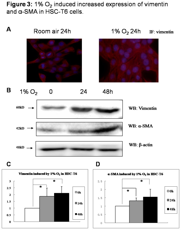
Figure 3. 1% O2 induced increased expression of vimentin and α-SMA in HSC-T6 cells.

HSC-T6 cells were cultured in room air or in 1% oxygen. (A) Vimentin expression in HSC-T6 cells grown on the coverslips at 48 hours post-seeding were co-stained with anti-vimentin (red) and DAPI (blue) by immunocytochemistry. (B) Cells were collected at indicated time and cell lysates were subjected to detect vimentin and α-SMA. Densitometric analysis was performed using pooled data from three such experiments. Data are mean ± SD. (C) vimentin; (D) α-SMA. * : P<0.05.
