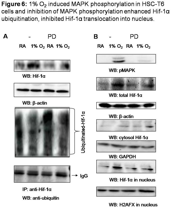
Figure 6. 1% O2 induced MAPK phosphorylation in HSC-T6 cells and inhibition of MAPK phosphorylation enhanced Hif-1α ubiquitination, inhibited Hif-1α translocation into nucleus.

Cells were pretreated with PD98059 (50µM) or vehicle (DMSO) for 1h, and then cultured in room air or in 1% oxygen at 37°C for 15min. (A) 50µg of cell lysates was subjected to detect Hif-1α and β-actin with western blot and 1mg of lysates was immunoprecipitated with anti-Hif-1α, followed by immunoblotting with anti-ubiquitin. (B) Cells were collected and protein samples extracted from total lysates, cytoplasm and nucleus were subjected to detect phosphorylated MAPK, Hif-1α, β-actin, GAPDH and H2AFX with western blot.
