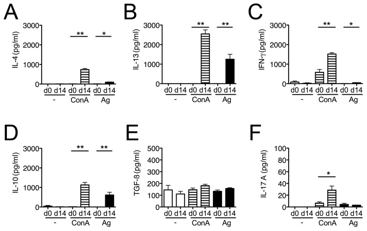
Figure 2. Cytokineproduction by mLN cells during acute H . p . bakeri infection.
IL-4 (A), IL-13 (B), IFN-γ (C), IL-10 (D), TGF-β (E) and IL-17A (F) were measured in mLN culture supernatants with (ConA, H.p.b-Ag) or without (-) stimulation. Cells were derived from naïve controls and mice at day 14 post infection. Mean + SEM is shown for 5 mice per group. H.p.b. Ag: H. p. bakeri antigen; Con A: concanavalin A; * p < 0.05; ** p < 0.005.

