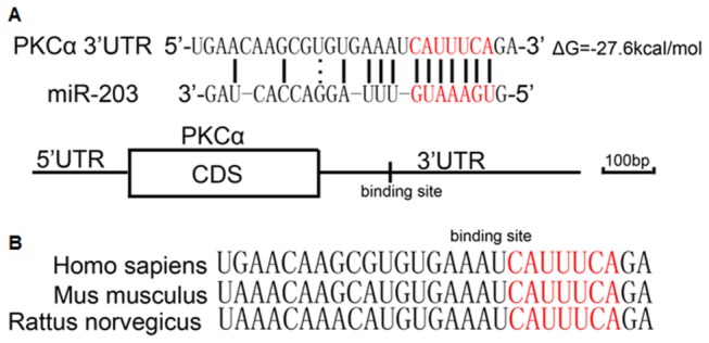
Figure 1. Identification of the conserved miR-203 binding sites within the PKCα mRNA 3’-UTR.

A, schematic description of the hypothesized duplexes formed by the interactions between the PKCα 3’-UTR binding sites and miR-203. The predicted structure of the base-paired hybrid is diagrammed. Paired bases are indicated by a black line, and G:U pairs are indicated by three dots. The predicted free energy of the hybrid is indicated. B, sequence alignment of the putative miR-203 binding sites across species. The seed complementary sites are marked in red, and all nucleotides in the regions are conserved in several species, including human, mouse and rat.
