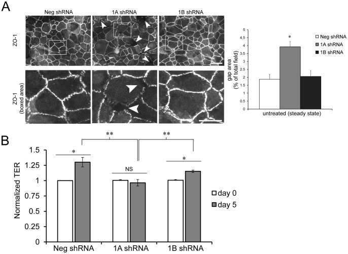
Figure 1. Rap1A knockdown in vitro impairs steady state monolayer integrity and decreases transepithelial electrical resistance.
(A) RPE cells transduced with indicated shRNA constructs were grown on coverslips for 4–5 days to obtain steady state monolayers. Top row, representative immunofluorescence localization of the junctional marker ZO-1. Scale bar = 50 µm. Boxed areas are enlarged to highlight intercellular gaps in Rap1A shRNA cell monolayers (bottom row). Scale bar = 25 µm. Quantification of total monolayer gap area per image field (expressed as % of total field area) using image analysis software (ImageJ) was used as an indication of monolayer integrity. Graph shows the mean of n = 4 fields ± SEM, representative experiment from 2 independent trials. **p<0.01, 1A shRNA compared to both Neg and 1B shRNA. (B) TER was measured on cells prior to shRNA knockdown (day 0), and again after 5 days. Graph shows TER of negative control, Rap1A, or Rap1B shRNA treated cells, representing average TER normalized to control at t = day 0 from n = 4 independent experiments. Control shRNA and Rap1B shRNA cells increased TER after 5 days in culture, while Rap1A shRNA cells did not significantly increase TER from day 0 to day 5. * p≤0.01, NS = not significant; **p≤0.01, TER of Rap1A shRNA monolayers significantly lower at steady state (day 5) compared to control and Rap1B shRNA.

