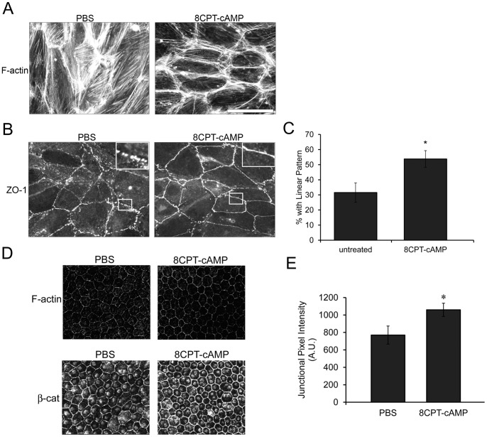
Figure 4. Activation of total Rap1 enhances recruitment of junctional proteins and cortical F-actin both in vitro and in vivo.
(A) 8CPT-cAMP treatment (250 µM, 1 hr) of cultured RPE monolayers decreases stress fibers and enhances cortical F-actin morphology. (B) Enhanced recruitment and linear junctional staining of ZO-1 with 8CPT-cAMP treatment compared to PBS control. Boxed areas are enlarged in the upper right inset to highlight differences in junctional staining pattern. Scale bar, 50 µm (C) Quantification of in vitro 8CPT-cAMP treatment. Percent of cells that show enhanced (linear) junctional localization of ZO-1 (average % of 10 fields/condition (>600 cells total counted). * p<0.01 (D) In vivo, eyes injected with 8CPT-cAMP also have enhanced linear junctional recruitment of proteins such as F-actin and β-catenin. (E) Quantification of junctional β-catenin in PBS vs. 8CPT-cAMP-injected eyes. Data plotted as average junctional pixel intensity from random cells per field, from n = 4 injected eyes (>100 cells). * p = 0.0324 compared to PBS-injected.

