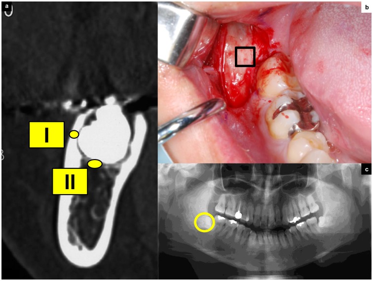
Figure 1. Design of the tooth extraction procedure and CT measurement.
(a) This CT image details the condition of third molar teeth covered with cortical bone. The regions of interest in CT image analysis were (I) the buccal cortical bone area, which was taken for biopsy during surgery; and (II) the region under the cortical bone, which indicates cancellous bone near the third molar tooth. (b) Removal of the third molar tooth. The envelope flap is raised, revealing the cortical bone. The line indicates the biopsy area. (c) Orthopantomographs showing impacted third molar teeth.

