
Figure 2.
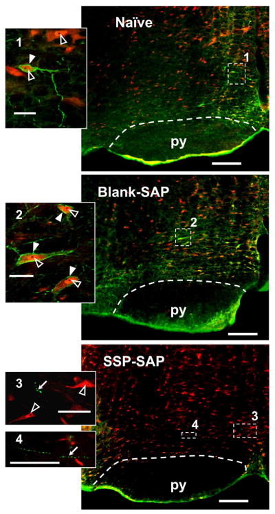
Examples of NK-1R labeling in single RVM sections (left side) from a naïve rat (top), and from rats pretreated with Blank-SAP (middle) and SSP-SAP (bottom). These single virtual tissue images were taken with a 10× objective. NK-1R-ir appears green and NeuN-ir appears red. In the RVM of naïve and Blank-SAP treated rats, labeling of NK-1R-ir neurons are similar. Pretreatment with SSP-SAP nearly abolished NK-1R-ir and few NK-1R-ire axons with varicosities could be found as a result of internalization of SP receptors (Mantyh et al., 1997). Inserts are single optical sections taken at 40× objective of the areas marked on the corresponding RVM by the numbered white dashed rectangles. NeuN-ir cell bodies with negative nucleoli (black) and NK-1R-ir plasma membrane are indicated by white arrowheads. NK-1R negative neurons with NeuN-ir nuclei with nucleoli are indicated by open arrowheads. NK-1R-ir varicosities induced by SSP-SAP are indicated by white arrows. Scale bars for the RVM and the inserts are 200 and 20 μm, respectively. Pyramidal tracts (py) are indicated by white dashed lines.