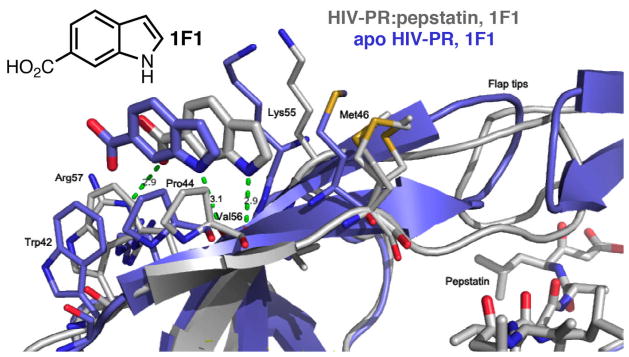
Figure 1.

Structure of HIV PR:pepstatin bound to 1F1 (gray) compared to structure of apo HIV PR bound to 1F1 (blue). Note the inverted orientation of the flap tips and the shift of the binding site for the apo-PR complex. Met46 is disordered in the PR:pepstatin structure and adopts three conformations with partial occupancy.
