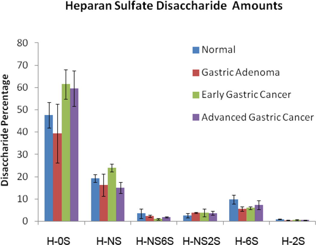
Figure 3.
Measured levels of HS disaccharides in GAGs present in stomach tissues. The HS disaccharide amounts, calculated by comparison to HS disaccharide standards, were normalized to the dry tissue weight of each sample. No significant differences were seen between the different stomach tissue types.

