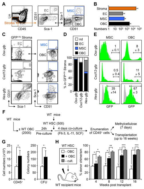
Figure 1. HSC-supportive activity of endosteal OBCs.
(A) Flow cytometry approach used to identify endosteal BM stromal populations. (B) Average numbers of ECs, MSCs and OBCs contained in the endosteal (Lin−/CD45−) BM stromal fraction of wild type (WT) mice (n = 23 in 7 independent experiments). (C) Immunophenotype and (D) frequencies of GFP+ endosteal ECs, MSCs and OBCs in Osx-gfp, Cxcl12-gfp and Nes-gfp reporter mice (n = 2–4 per genotype; nd: not determined). (E) Frequency of GFP+/hi cells in endosteal MSCs and OBCs of Osx-gfp, Cxcl12-gfp and Nes-gfp reporter mice (green histograms). Grey histograms indicate background GFP fluorescence levels in control populations. (F) Schematic of the short-term co-culture of HSCs with or without OBCs, and follow up analyses. (G) Cell numbers and methylcellulose colony-forming unit (CFU) activity. (H) Transplantation in lethally irradiated WT CD45.1 recipients (n = 3–5 mice per group, with results replicated in another independent experiment). Mice were bled every 4 weeks and analyzed for the percentage of CD45.2+ donor-derived cells (- cult.: no culture).
Data are means ± SD; *p ≤ 0.05, ** p ≤ 0.01, ***p ≤ 0.001. See also Figure S1.

