
Figure 2.
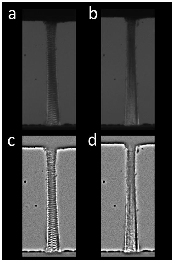
(a) Tendon imaged with the polariscope prior to loading. Crimp bands visible throughout the tissue. (b) At 4% strain, crimp bands disappear. (c–d) Bandpass-filtered tendon images before and after stretching to 4%. Crimp bands are easier to distinguish in the processed images.