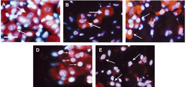
Fig 1.

Morphological features of apoptosis in sensory neurons. Propidium iodide (red) and Hoechst 33342 (blue) staining revealed morphological changes of apoptosis in dorsal root ganglion (DRG) sensory neurons. A. normal sensory neurons with large cell body and nucleus from freshly prepared DRG (0 hour). Sensory neurons after 24 (B), 48 (C), 72 (D) and 96 hours (E) displayed cell shrinkage as well as nuclear and chromatin condensation. Scale bar: 25 µm. Arrows show sensory neurons.
