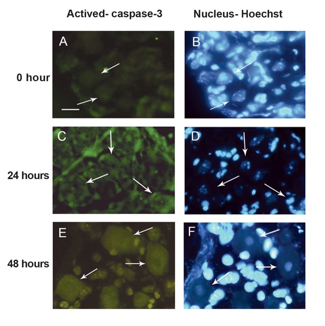
Fig 4.
: Immunolocalization of activated caspase-3 antibody in dorsal root ganglia (DRG) sensory neurons. Sensory neurons were stained with activated caspase-3 antibody (green) and counterstained with Hoechst 33342 (blue). (A-B).Weak activated caspase-3 immunoreactivity in sensory neurons from DRG at 0 hour with no sign of apoptotis. Sensory neurons from DRG cultured for 24 hours (C-D) and 48 hours (E-F) displayed intense activated caspase-3 immunoreactivity both in the nucleus and the cytoplasm where the nuclei showed apoptotic features. Scale bar: 25 µm. Arrows show sensory neurons.

