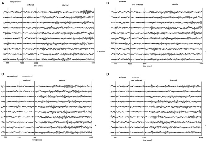
Figure 3.
Raw LFP traces (1–475 Hz) during PA (A,B) and BFS (C,D) for 10 trials from a typical prefrontal recording site. In (A), a non-preferred stimulus is presented in one eye and after 1 s is removed and a disparate pattern is presented in the contralateral eye. Using as a criterion the discharge response of the locally recorded population we determined that the second stimulus was the “preferred.” In (B) the order of visual stimulation is reversed and the preferred stimulus is followed by the presentation of the non-preferred. In (C,D) for BFS the order of stimulation is the same as in (A,B), respectively. However, in these trials the stimulus presented first is not removed but remains on and is suppressed by the stimulus presented between t = 1301–2300 ms (dominant stimulus-black, suppressed stimulus-gray). In both PA and BFS and for all conditions we can observe that the onset or change of visual stimulation results in a remarkable suppression of low frequency-high amplitude LFP components that rebound later when the stimulus remains on. These components are particularly dominant during the inter-trial period (t = 1301–2300).

