Figure 2.
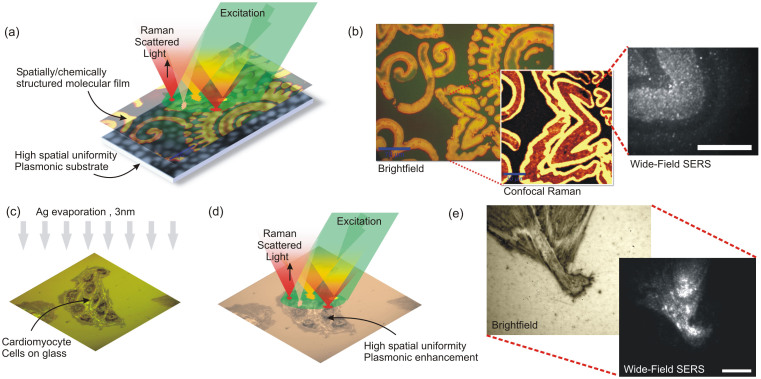
(a) A highly uniform plasmonic substrate can be used for confocal or wide-field Surface Enhanced Raman Spectroscopic (SERS) imaging of biological architectures. (b) AIPs imprinted on the substrate can be observed in bright-field images, due to the contrast generated by a shift of the plasmonic absorption band. Uniformity of the surface allows confocal Raman mapping of the peptide overlayer. Under wide-field excitation (scale bar 10 μm), blinking spots correlated with the presence of peptides can be resolved (see Supplementary Video 1). (c) Cardiomyocytes are covered with 3 nm Ag, which segregate into nanoislands with 20 nm average diameter. (d) Thin Ag overlayer provides high-spatial uniformity plasmonic enhancement. (e) Brightfield and wide-field SERS images (scale bar 10 μm) show correlation of blinking spots with the presence of organic material from the cells (see Supplementary Video 3).

