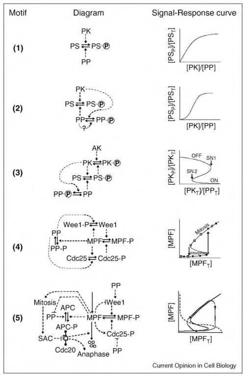
Figure 1.
Regulatory motifs. In each motif, a solid arrow indicates a chemical reaction, and a dashed arrow indicates an enzyme that catalyzes the reaction. (1) The basic motif. A protein substrate (PS) is phosphorylated by a protein kinase (PK) and dephosphorylated by a protein phosphatase (PP). The incoming ‘signal’ is the ratio of PK activity to PP activity; and the response is the fraction of PS in the phosphorylated form. The response saturates at f = 1 for large values of the PK:PP ratio. [PST] = [PS] + [PSP] = total concentration of the substrate. (2) Coherent feed-forward loop. PK phosphorylates and inactivates PP. The active form of PP may promote its own accumulation by auto-dephosphorylation (the dotted feedback signal marked with a ‘?’). The signal–response curve is now sigmoidal. (3) Positive feedback loop. The protein substrate of motif #2 is now a phosphatase that activates PK by reversing an inhibitory phosphorylation carried out by the ‘antagonistic kinase’ (AK). In this case, the response variable, the fraction of PK in the phosphorylated form, corresponds to the low-activity state of PK. The signal–response curve shows a region of bistability between the points marked SN1 and SN2. (4) Bistability in the activation of MPF. MPF (mitosis promoting factor) is a heterodimer of cyclin B and Cdk1. Wee1 phosphorylates MPF on an inhibitory residue of the Cdk1 subunit. Cdc25 removes the inhibitory phosphate group. PP2A is the phosphatase that opposes the phosphorylation of Wee1 and Cdc25 by MPF. In the signal–response curve, [MPFT] = [MPF] + [MPFP] = total MPF = total concentration of cyclin B, because the Cdk1 subunit is present in excess and free Cdk1 molecules have no kinase activity. The signal–response curve shows a robust region of bistability for intermediate levels of total cyclin B. (5) Negative feedback and oscillations. The positive feedback loops of motif #4 are supplemented by a negative feedback loop, whereby MPF activates the APC/Cdc20 complex, which initiates the proteolysis of cyclin B. The dashed lines from MPF to Wee1, Cdc25, and so on, indicate that MPF has an effect (activation or inhibition) on the target protein, without specifying the precise molecular mechanism of this effect. This is a shorthand convention to simplify the diagram, and the mechanism of the effect can be deduced from previously described motifs. The ‘dumbbell’ notation indicates the reversible formation of a complex between proteins A and B at the ends of the dumbbell, and an icon for the complex (overlapping white rectangles) is placed on the middle of the dumbbell. Four small circles indicate degradation products of a protein. On the signal–response plane, there is no stable steady state, and the system executes periodic oscillations around the trajectory indicated by the dotted line.

