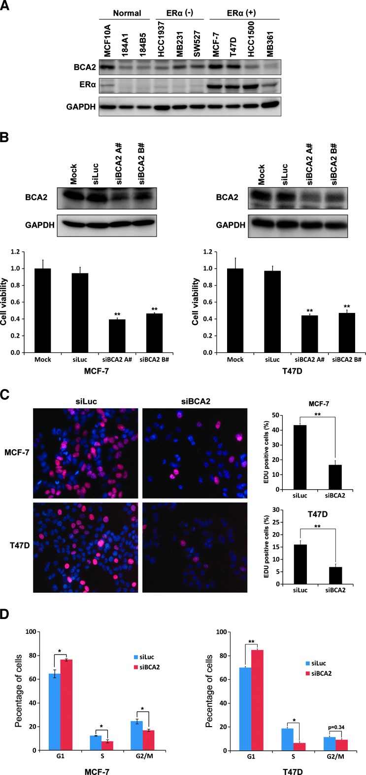
Figure 1.
Knockdown of BCA2 inhibits breast cancer cell proliferation, DNA synthesis, and cell cycle. (A) WB analysis of BCA2 protein levels in a panel of immortalized breast epithelial cell lines and ERα-negative and ERα-positive breast cancer cell lines. GAPDH was used as a loading control. (B) MCF-7 and T47D cells were transfected for 72 hours with either Lipofectamine 2000 reagent as mock, control siLuc RNA, siBCA2-A#, or siBCA2-B#. The knockdown effect was assessed by WB analysis, and cell viability was examined using the SRB assay (**P < .01 compared to siLuc). (C) After silencing of BCA2 by siBCA2-A#, MCF-7 and T47D cells were pulse labeled with EdU Alexa Fluor 647 for 4 hours and were stained the nuclei with 10 µg/ml PI. The percentage of positive cells incorporated with EdU were shown in the histograms (right side, **P < .01). All quantitative values were from triplicate experiments. (D) After silencing of BCA2 by siBCA2-A#, MCF-7 and T47D cells were harvested, fixed, stained with PI solution, and analyzed by flow cytometry. The percentage of arrest G1 phase cells was shown in the histograms (*P < .05 and **P < .01).

