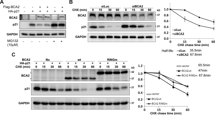
Figure 3.
(A) BCA2 promotes p21 protein degradation through proteasome. Flag-BCA2 and HA-p21 were cotransfected into HEK293FT cells as indicated. The proteasome inhibitor MG132 (10 µM) was added to treat the cells for 8 hours if necessary. (B) After depletion of BCA2 by siBCA2-A# in MCF-7 cells, the cells were exposed to CHX for a time as indicated, and WB analysis was performed. The quantitative half-lives of p21 protein from two independent experiments are shown on the right side. (C) HEK293FT cells were cotransfected with HA-p21 and WT BCA2 or RING finger mutant BCA2 expression plasmids. The BCA2-RINGm protein migrates much slower than WT BCA2 possibly because of the protein structure difference under the electrophoresis condition. The cells were exposed to CHX for a time as indicated. The level of p21 protein was detected by WB analysis. The quantitative data from two independent experiments are shown on the right side.

