
Figure 3.
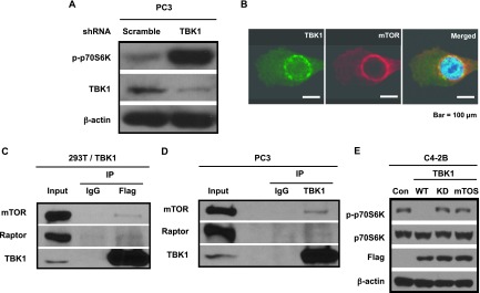
TBK1 inhibits mTOR signaling. (A) PC3 cells were infected with lentivirus containing scrambled or TBK1 shRNA plasmid, selected with puromycin, and analyzed by Western blot analysis using antibodies against p-p70S6K, TBK1, or β-actin. (B) PC3 cells were co-immunostained with TBK1 (green) and mTOR (red) antibodies. Merged image (yellow) shows co-localization of TBK1 and mTOR. (C) 293T cells were transfected with a plasmid encoding Flag-TBK1. Cell lysates were immunoprecipitated with Flag antibodies or normal mouse IgG as negative control and analyzed by Western blot analysis using antibodies against mTOR, raptor, and TBK1. (D) PC3 cell lysates were immunoprecipitated with TBK1 antibodies or normal rabbit IgG as negative control and analyzed by Western blot analysis using antibodies against mTOR, raptor, and TBK1. (E) C4-2B cells were transfected with control empty vector (Con) or a plasmid encoding wild-type (WT) or Kinase-dead (KD) or TOS motif mutant TBK1 (mTOS). Cell lysates were analyzed by Western blot analysis using antibodies against p-p70S6K, p70S6K, Flag, and β-actin.