
Figure 5.
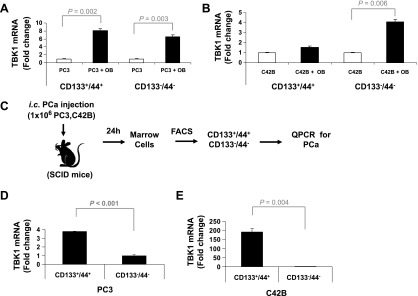
TBK1 is highly expressed in PCa stem-like cells on mouse BM niche. (A) PC3 or (B) C42B cells were sorted with APC-anti-CD44 and PE-anti-CD133 antibodies. The CD133+/CD44+ and CD133-/CD44- cells were cultured for 48 hours on tissue culture plastic or ST2 (OB) cells. Expression of TBK1 was quantified by qPCR. The results were normalized to β-actin. (C) Experimental model of isolation of PCa stem-like cells in vivo. The CD133+/CD44+ and CD133-/CD44- cells were sorted from BM cells of (D) PC3- or (E) C4-2B-injected SCID mice. Expression of TBK1 was quantified by qPCR. The results were normalized to β-actin. All results represent average values from triplicate assays, and the experiments were performed twice.