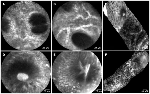
Figure 2.

Colonic mucosa. A: Patient in remission from ulcerative colitis. Crypt distortion and fusion and many capillaries visible in the lamina propria; B: Patient in remission from ulcerative colitis. Enlarged spaces between crypts and dilated prominent branching vessels; C: In patient with active ulcerative colitis (distal colitis). Image of colonic mucosa showing the switch from normal mucosa (top of the figure) to inflamed mucosa Inflamed mucosa showing irregular arrangement of crypts, crypt fusion and capillaries alterations; D: In patient with active ulcerative colitis. Dilated and bright crypt lumen (fluorescein leakage) with intact epithelium; E: In patient with active ulcerative colitis. Dilated, irregular and bright crypt lumen (fluorescein leakage) with partially intact epithelium; F: In patient with highly active ulcerative colitis (Mayo CU3). Crypts distortion and destruction, crypt abscess and crypts replacement by diffuse necrosis.
