


Figure 2.
FBXL4 Protein Structure and Localization
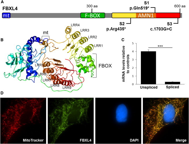
(A) Schematic of FBXL4 showing the location of the bioinformatically determined mitochondrial localization sequence (mt) and conserved domains (F-box CDD ID pfam12937, AMN1 CDD ID cd09293). The locations of each of the mutations are indicated.
(B) Three-dimensional structural modeling of FBXL4 shows the canonical folds of the F-box and nine leucine-rich repeats. The protein schematic and 3D structure are both displayed in reverse rainbow from the N to C terminus.
(C) Splicing of exon 9 was demonstrated to be significantly impaired in S3 compared to controls. Error bars indicate standard deviation; ∗∗∗p < 0.0001.
(D) FBXL4 was confirmed to be a mitochondrial protein through an immunolocalization study in human fibroblasts, showing HA-tagged FBXL4 (green) colocalizing with MitoTracker Red-labeled mitochondria. Nuclei were counterstained with DAPI (blue).