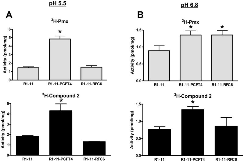
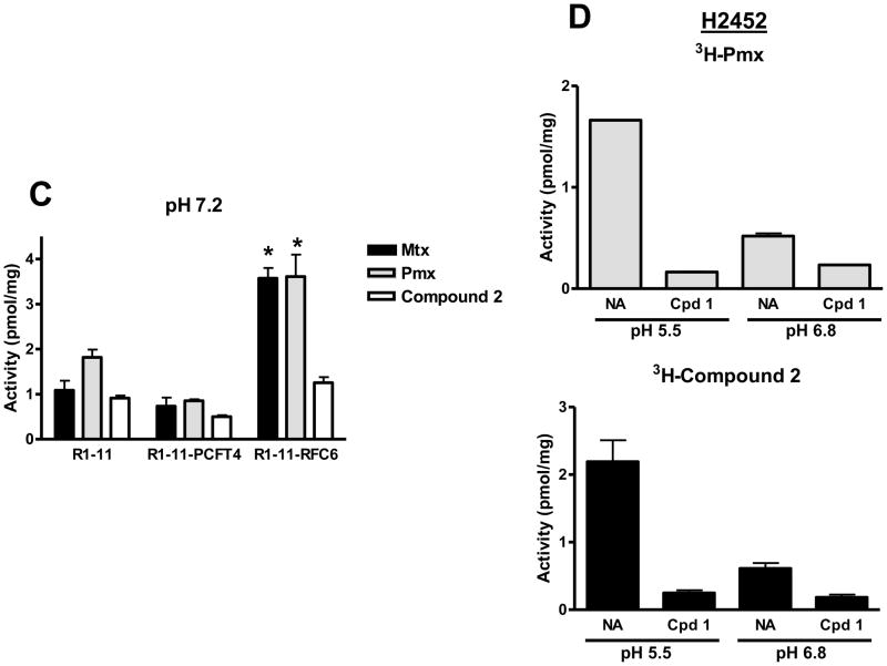
Figure 6. pH-dependent transport of Pmx and compound 2 in R1-11 HeLa and H2452 MPM sublines.
Panels A and B: Cellular uptake was measured over 5 minutes in R1-11 mock (labeled R1-11), R1-11-PCFT4, and R1-11-RFC6 HeLa cells at 37°C with [3H]Pmx and [3H]compound 2 (both at 0.5 μM) in pH 5.5 (panel A) and pH 6.8 (panel B) buffers. Details are provided in Materials and Methods. In panel C, cellular uptakes of [3H]Mtx, [3H]Pmx, and [3H]compound 2 (each at 0.5 μM) were measured over 5 minutes at pH 7.2 in anion-free Hepes-sucrose-Mg2+ buffer. For panels A–C, statistically significant differences (p < 0.05) in [3H]Pmx or compound 2 uptake levels between R1-11-PCFT or R1-11-RFC6 and those in R1-11 cells are noted (*). In panel D, cellular uptake was measured over 5 min in H2452 MPM cells for [3H]Pmx and [3H]compound 2 (both at 0.5 μM) at pH 5.5 and pH 6.8 in the absence and presence of 10 μM unlabeled compound 1. For both [3H]Pmx and [3H]compound 2, differences in the absence


