FIG. 1.
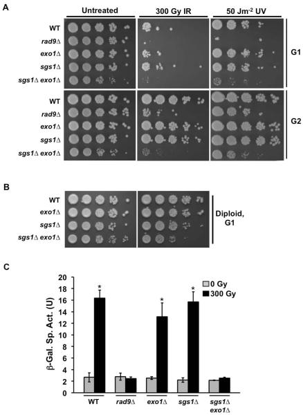
G1-arrested sgs1Δ exo1Δ mutants are sensitive to DNA damage. (A) Viability of haploid cells exposed to DNA damaging agents during G1 and G2 phases. Spot assays were performed on G1- and G2-arrested cells. Overnight cultures were diluted to 0.2 OD600 and arrested in G1 or G2 with αF (10 μM) and nocodazole (15 μg ml−1) respectively. Ten-fold serial dilutions were plated on solid media and exposed to IR (300 Gy), UV (50 Jm−2), or mock-treated. Plates were incubated at 30°C for 2 days prior to image capture. (B) Viability of diploid cells exposed to DNA damaging agents during G1 phase. Overnight cultures were diluted to 0.2 OD600 and arrested in G1 by incubation for 4 hrs in media lacking a nitrogen source. Ten-fold serial dilutions were plated on solid media and exposed to IR (300 Gy) or mock-treated. Plates were then incubated at 30°C for 2 days prior to image capture. (C) Activation of RNR3 promoter in response to DNA damage response (DDR) in G1 cells. Overnight cultures were diluted and arrested in G1 using αF. Cells were mock treated or exposed to 300 Gy IR. 45 mins after irradiation, protein samples were extracted and assayed for β-galactosidase activity by spectroscopic measurement of cleaved ONPG product. A measurement of the β-galactosidase activity is shown. Error bars represent standard deviation from three independent experiments (*, P<0.05).

