
Figure 2.
Pathogenic Variants of HCFC1 in cblX
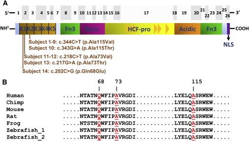
(A) The top panel shows the 26 exons of HCFC1 as gray boxes. The bottom panel shows the predicted HCFC1 domains, including the kelch domain (kelch motifs K1–K5), Fn3 (fibronectin type 3), the basic domain, HCF-proteolysis repeats (HCF-pro; represented as triangles), the acidic domain, and NLS (nuclear localization signal) domains (adapted from Wilson et al.24). The HCFC1 mutations are clustered within exon 2 and 3 of the cDNA, corresponding to the first (K1) and second (K2) kelch motifs, respectively, in HCFC1.
(B) Comparative analysis of HCFC1 from multiple species demonstrated that Gln68, Ala73, and Ala115 (highlighted in red) are evolutionarily conserved throughout vertebrates.