
Figure 4.
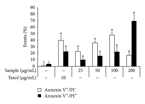
Simultaneous in vitro detection of apoptosis (annexin V staining) and necrosis (PI staining) of 4T1 cells treated with P. dentata F2 fraction. The 4T1 cells were incubated in DMEM culture containing 10% FBS and F2 sample at indicated concentrations for 48 h. Taxol (10 μg/mL) was used as a positive control. The relative cell population (%) undergoing a solely apoptotic (annexin V positive) pathway is shown by an open bar square; cells undergoing necrotic-apoptotic pathway (annexin V and PI double positive) are shown by a closed black bar square. Data are expressed as mean ± S.D. (n = 3).