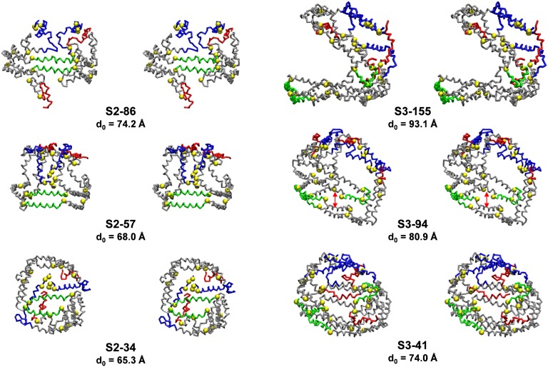
Fig. 8.
Cross-eyed stereo licorice images of protein conformations of the S2 and S3 particles after CGMD simulations. The three S2 particles (left-hand column) contain two apoA-I in a double belt, and the three S3 particles (right-hand column) contain three apoA-I in a double belt plus hairpin. From top to bottom, the S2 and S3 particles show the effects of core and surface lipid reductions on protein conformation. Residues 1–43, blue: helix 5, green; helix 10, red; prolines, yellow space filling. The unhydrated diameters calculated from volumes of lipid and protein components assuming a perfectly spherical shape is designated d0.

