
Figure 2.
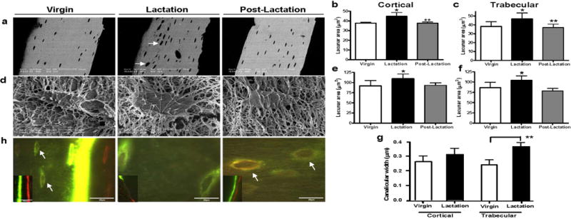
Lactation induces osteocytic remodeling during lactation that returns to virgin levels with postlactation. (A) BSEM showed that during lactation osteocyte lacunae enlarged with irregular borders indicating mineral removal (white arrows). (B) Osteocyte lacunar area (in μm2) was significantly increased in cortical bone during lactation as compared to virgin or 7 days postlactation. (Experiment repeated four times; *p < 0.05; **p < 0.01; n = 7–9 per group). (C) Osteocyte lacunar area (in μm2) was significantly increased in trabecular bone during lactation as compared to virgin or 7 days postlactation (*p < 0.05; **p < 0.01; n = 7–9 per group). (D) Osteocyte lacunae were imaged using acid-etch resin-casted SEM to further validate the measurements by SEM. (E) Osteocyte lacunar area measured by acid-etch resin-casted SEM was significantly increased in cortical bone during lactation as compared to virgin or 7 days postlactation (*p < 0.05; n = 4–5 per group). (F) Osteocyte lacunar area measured by acid-etch resin-casted SEM was significantly increased in trabecular bone during lactation as compared to virgin or 7 days postlactation (*p < 0.05; n = 4–5 per group). (G) The same resin-casted SEM images were also used to measure canalicular diameter. An increase in canalicular diameter was observed in cortical bone from lactating animals and a significant increase in trabecular bone (p < 0.001; n = 4–5 per group). (H) Double fluorochrome labeling. Two distinctive lines of fluorochrome labeling, calcein green (first injection) and Alizarin red (second injection), can be found at the bone surface (smaller image insert). In the virgin animal, faint label was taken up by the newly embedding osteocytes at the mineralization front (arrows in virgin). Only green labeling was observed in the lactating animals and an intermittent green label on the bone surface. Lacunae labeled with both fluorochromes (mixed green and red bands) were observed distant from the mineralization front (previously existing lacunae) in the postlactation animals (white arrows in postlactation).