
Figure 6.
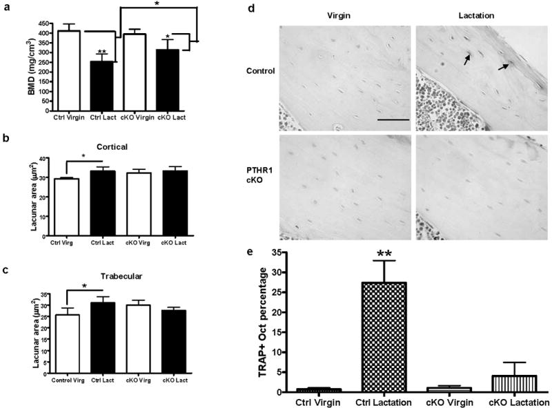
Osteocyte remodeling during lactation was blocked in osteocyte-specific PTHR1-cKO mice. (A) BMD measurement by μCT showed a decrease in bone density in both the control and the PTHR1-cKO mice during lactation. There is less bone density loss (47% attenuation) during lactation in PTHR1-cKO mice (*p < 0.05; **p < 0.01; n = 3–6 per group). (B) In control mice, osteocyte lacunar area in tibial cortical bone significantly increased during lactation compared to virgin, while lacunar area did not enlarge in the PTHR1 cKO mice with lactation (*p < 0.05; n = 3–6 per group). (C) In control mice, osteocyte lacunar area in tibial trabecular bone significantly increased during lactation compared to virgin, while lacunar area did not enlarge in the PTHR1 cKO mice with lactation (*p < 0.05; n = 3–6 per group). (D) Tartrate resistant acid phosphatase staining. A significant increase in TRAP-positive osteocytes in control mice with lactation was not observed in the PTHR1-cKO mice. (The periosteal surface is upper right and endocortical surface is lower left.) (E) In control mice, a significantly increased number of osteocytes with TRAP activity (p < 0.01) in the lactating mice (27.4% ± 9.6%** TRAP+ osteocytes) was observed compared to the virgin mice (0.7% ± 0.9%). There was no significant difference in TRAP activity from PTHR1-cKO mice during lactation (4.1% ± 8.4%) compared to virgin mice (1.1% ± 1.2%) (*p < 0.05; **p < 0.01; n = 3–6 per group).