
Figure 7.
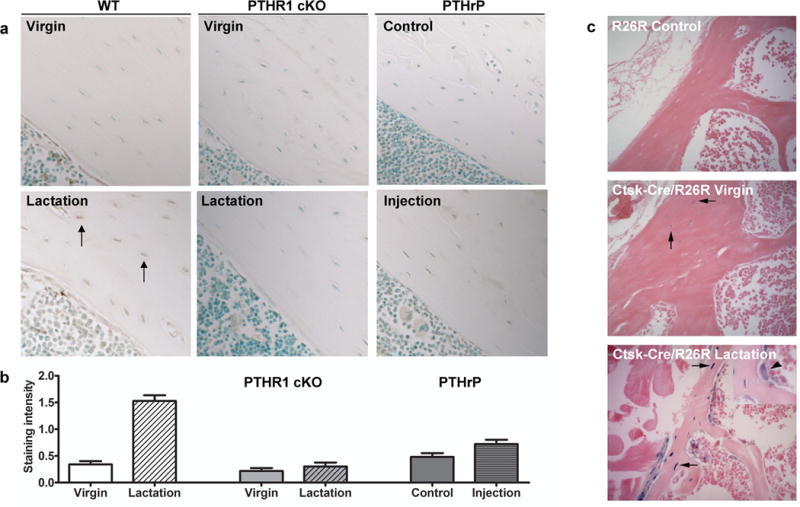
Cathepsin K protein and gene expression is elevated in osteocytes during lactation. (A) Immunostaining for cathepsin K in osteocytes (black arrows), scale bar 100 μM. Increased staining is observed in bone from wild-type lactating animals compared to virgin animals. No obvious increases were observed in PTHR1 cKO virgin or lactating animals, nor significant differences with PTHrP-injected animals compared to vehicle control–injected animals. (B) Immunostaining was quantitated on all groups within the same experiment showing significant increases in lactating wild-type animals compared to the other groups. (C) Bone from virgin mice containing both the Ctsk-cre and the R26R showed some pale blue staining in osteocytes (arrows, middle panel), whereas bone from lactating animals showed that not only osteoclasts, but more osteocytes express LacZ and thus cathepsin K. The arrowhead (inset, lower panel) indicates the osteoclast as a positive control for beta-galactosidase staining. The R26R control bone (upper panel) shows no blue color, as expected.