
Figure 1. MRIs of patient with primary CNSL.
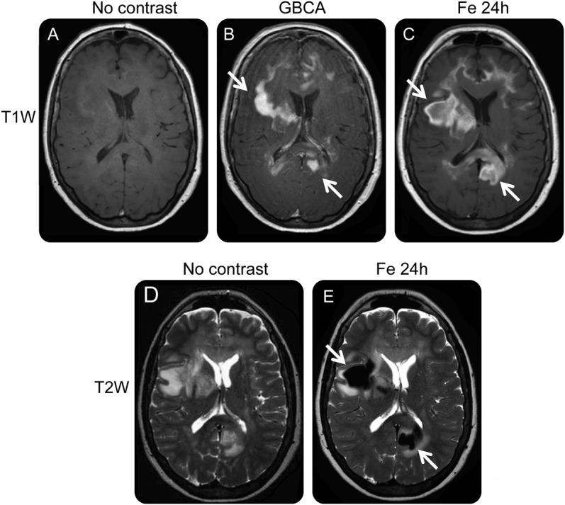
MRIs from case 1. (A) T1-weighted MRI without contrast agent. (B) T1-weighted MRI immediately after administration of GBCA shows multifocal, enhancing, deep white matter lesions (arrows) suggesting primary CNSL. Note, however, that the relatively minimal mass effect for lesion extent and incomplete enhancing rims are more typical of demyelinating disease. (C) T1-weighted MRI 24 hours after administration of USPIO contrast shows more extensive enhancement because of phagocytic cellular uptake (arrows) than the GBCA-enhanced scan. (D) T2-weighted MRI without contrast shows multifocal confluent areas of T2 hyperintensity in white matter. (E) T2-weighted MRI 24 hours after ferumoxytol shows multiple areas of intense USPIO uptake (arrows). CNSL = CNS lymphoma; GBCA = gadolinium-based contrast agent; USPIO = ultrasmall superparamagnetic iron oxide.