Fig. 1.
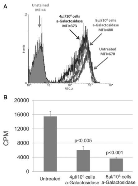
(A) Gal expression on untreated and α-galactosidase-treated WT porcine aortic endothelial cells (pAECs) was measured by flow cytometry. The mean fluorescence (MFI) was reduced from 670 (untreated) by 28% to 480 by a treatment of 4 μl/106 cells α-galactosidase and by 44% to 373 at 8 μl/106 cells. For reference, the average MFI from all GTKO pAECs was five (not shown), and the MFI of unstained cells was four. (B) Human peripheral blood mononuclear cell (PBMC) proliferation was reduced following treatments of WT pAECs with α-galactosidase. In MLR, WT pAECs treated with α-galactosidase at 4 and 8 μl/106 reduced Gal expression by 28 and 44% and stimulated 61 and 77% less human PBMC proliferation, respectively, than those untreated. PAEC viability was confirmed before MLR and equal numbers of stimulators and responders were used in each study. 3H incorporation values are presented as counts per minute. Data represent the mean plus or minus standard error of the mean (±SEM) and are representative of three different experiments.

