
Fig. 3.
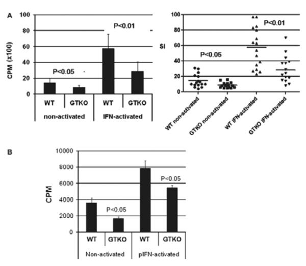
(A) The proliferative response of human peripheral blood mononuclear cells (PBMCs) (n = 15) to WT and GTKO porcine aortic endothelial cells (pAECs) before and after activation by pIFN-γ. This response was significantly less to GTKO pAECs before (P < 0.05) and after (P < 0.01) activation. PBMC proliferation is presented as counts per minute (CPM) for 3H incorporation (left) and in units of SI (right). Data represent the mean (±SEM) and are shown. (B) The proliferative response of baboon PBMCs (n = 4) to WT and GTKO pAECs before and after activation by pIFN-γ. The response to GTKO pAECs was significantly less before (P < 0.05) and after (P < 0.05) activation. 3H incorporation values are presented as CPM. Data represent the mean (±SEM) and are representative of three different experiments.