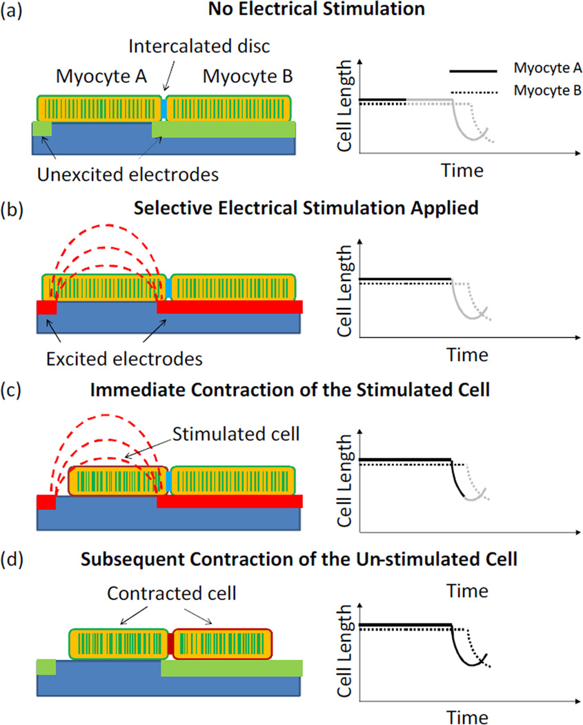
Fig. 1.
Schematics of the microengineered approach for intercellular communication studies. (a) A cardiac myocytes doublet was positioned in the electrodes array with one cell lying between two neighboring electrodes and the other one lying on top of an electrode finger. (b) Electrodes were selectively excited, giving rise to a localized electric field. (c) Myocyte A contracted upon the electrical stimulation. (d) Myocyte B contracted due to the electromechanical coupling with myocyte A through the intercalated disc. Left: Schematics of electrode stimulation and cell contraction; Right: Schematic curves showing the changes in cell length.

